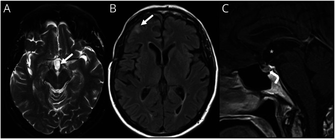
Figure 2. MRI Brain Postresection and Radiosurgery of the Adamantinomatous Craniopharyngioma.
Axial T2-weighted (A) and fluid-attenuated inversion recovery (B) MR brain images and sagittal postcontrast T1-weighted image (C) demonstrating postoperative changes after tumor resection and radiosurgery with resection cavity in suprasellar region (arrow, A; asterisk, C), gliosis in right frontal lobe beneath a craniotomy defect (arrow, B), resolution of hydrocephalus, and normal appearance to the pituitary gland (curved arrow, C).

