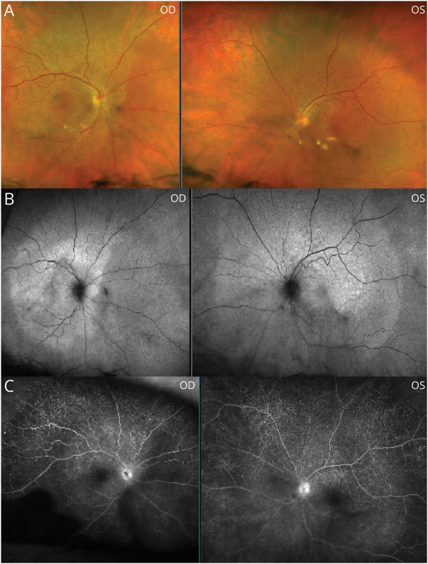
Figure 1. Fundus Photography, Fundus Autofluorescence, and Fluorescein Angiography.
Placoid-like macular pigmentary changes are depicted on fundus photography (A) and fundus autofluorescence (B); the sharply delineated margin of the pigmentary change (indicative of the pathologically affected area) is seen. Note also patchy choroidal filling and disc leakage noted on fluorescein angiography (C). OD = right eye; OS = left eye.

