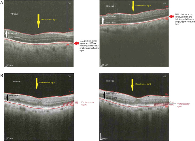
Figure 2. OCT Before Treatment and 14 Months Post-treatment.
This OCT (A) before treatment with intravenous penicillin demonstrates loss of the delineation of the outer retinal layers—the ELM, photoreceptor layers, and RPE layers—which can be visually differentiated from one another in the healthy eye—and are here supplanted by a single, hyper-reflective layer. These changes correlate with loss of the retina peripheral to the ellipsoid zone on OCT, specifically including the RPE and photoreceptor layers. This OCT (B) from 14 months post-treatment with intravenous penicillin now demonstrates visible delineation between the outer retinal layers, including the ELM, the photoreceptor layers, and the RPE. BM = Bruch membrane; ELM = external limiting membrane; ILM = internal limiting membrane; OCT = optical coherence tomography; RPE = retinal pigment epithelium.

