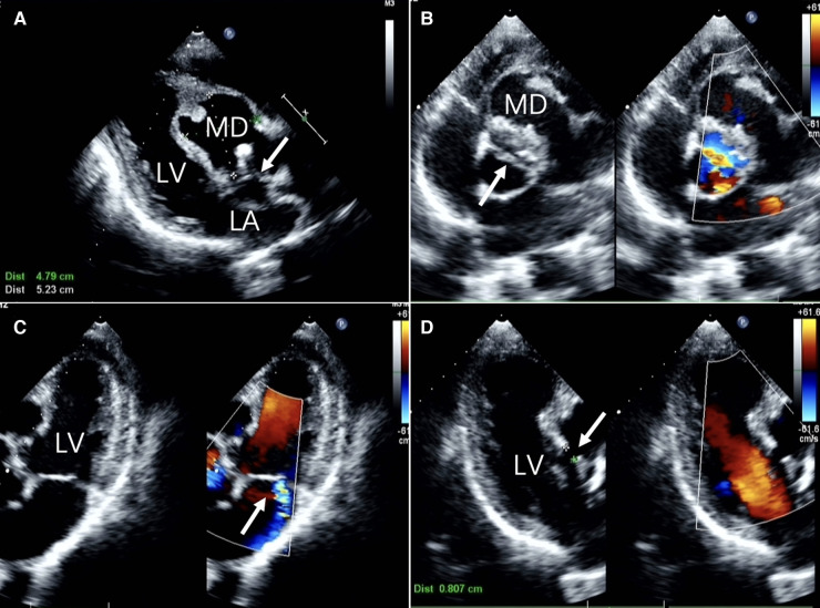
Figure 1.
Two-dimensional transthoracic echocardiographic images (A) long-axis view of the left ventricle showing echo interruption, no echo in the interventricular septum.(white arrow) (B) the short-axis cross-sectional view of the aorta demonstrates a bicuspid aortic valve anomaly. (white arrow) (C) the non-standard pentachamber cardiac section reveals mitral valve eccentric regurgitation. (D) The three-chamber cardiac cross-section reveals the dimension of the orifice. LA, left atrium; LV, left ventricle; MD, Myocardial dissection.

