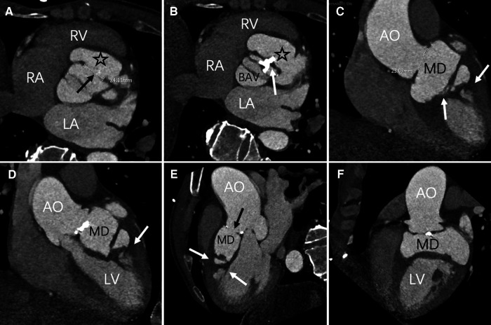
Figure 2.
Axial position, contrast-enhanced CT imaging reveals the maximum aperture of the sinus of Valsalva aneurysm to be 2.4 cm (black arrow). The myocardial dissection originates from the fused left and right coronary sinuses (asterisk). A bicuspid aortic valve anomaly is evident, with valve leaflet thickening and calcification (white arrow) (C–F) multiPlanar Reconstruction (MPR) exhibits irregular MD, fusion of the left and right coronary sinuses (black arrow), and high-density opacities within the ventricular wall and interventricular septum (white arrow). LA, left atrium; LV, left ventricle; RA, right atrium; RV, right ventricle; AO, Aortic; MD, Myocardial dissection; BAV, bicuspid aortic valve.

