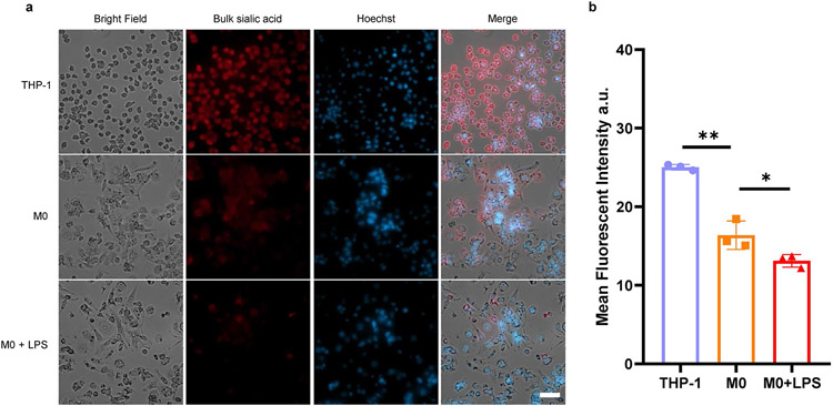
Extended Data Fig. 9 ∣. Fluorescence imaging of bulk sialic acid on the cell surface of THP-1 monocyte, resting M0 macrophage (M0), LPS activated M0 macrophage (M0 + LPS).
(a) Representative images of total sialic acid. These cells were metabolically labeled by Ac4ManNAz for 24 h, followed by incubation with DBCO-PEG4-biotin and Cy5-streptavidin for fluorescence imaging. Scale bars for cell image: 50 μm. (b) Quantification of the mean fluorescent intensity of the images, n = 3 biological replicates. Data are mean ±S.D. Unpaired two-tailed Student’s t-test determines the statistical significance. (**) p = 0.0013, (*) p = 0.0469.

