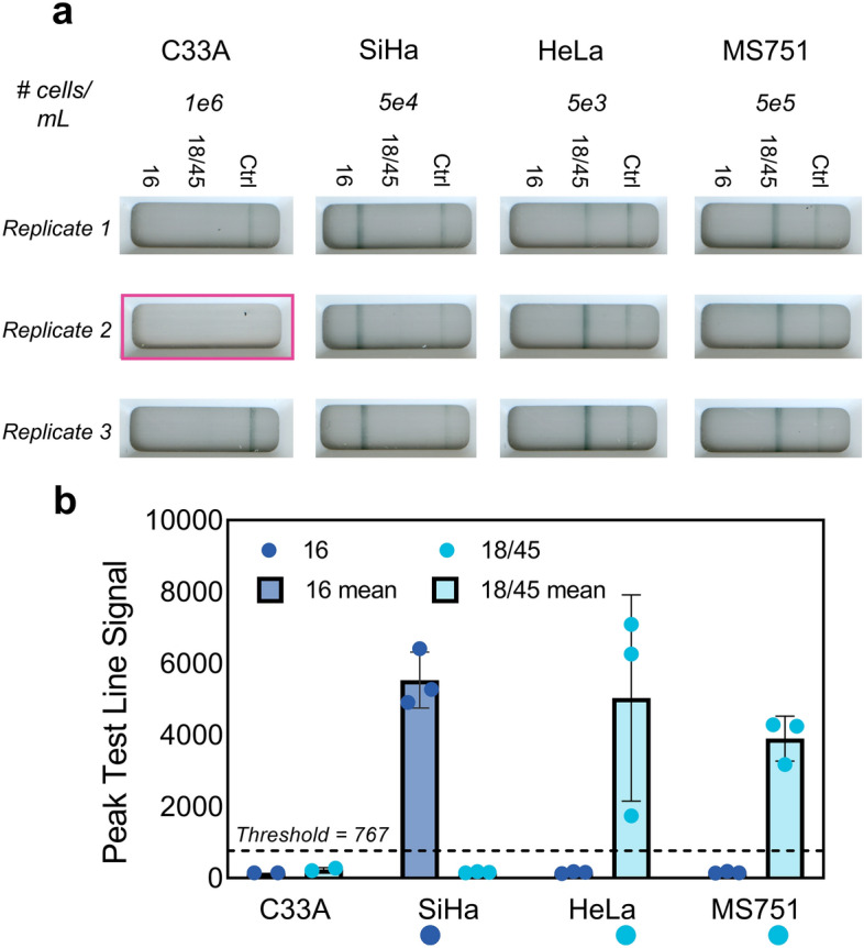
Figure 6.

Sample-to-answer testing with HPV-negative and HPV-positive cells. (a) C33A (HPV-negative), SiHa (HPV16-positive), HeLa (HPV18-positive), or MS751 (HPV45-positive) cells at the indicated concentrations were processed with a point-of-care compatible ACP-based sample preparation protocol, and the resultant lysates were amplified on NATflow instrumentation and detected in the integrated NATflow cartridge. Replicate cartridge images are shown (n = 3), with the highlighted cartridge image indicating an invalid test due to lack of fluid flow down the lateral flow strip. (b) Mean ± standard deviation of the peak test line signal intensity, as determined by the AX-2X-S lateral flow reader of the replicates shown in (a). Circles represent individual replicates, and circles beneath the x-axis indicate expected 16 or 18/45 positivity. All cellular lysates generated strong visible signal at the target HPV test line above the positivity threshold (dashed line) and had clean signals at the off-target test lines. C33A resulted in no signal generation at either the HPV16 or the HPV18/45 test line.
