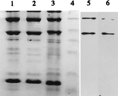
FIG. 3.
Profiles of phage structural proteins as determined by SDS–15% PAGE with the corresponding immunoblots for MAbs 2A5 and 6G7. Phage proteins were stained with Coomassie blue. Lane 1, phage Q44; lane 2, phage Q38; lane 3, phage c2; lane 4, prestained molecular mass markers (25, 32.5, 47.5, 62, 83, and 175 kDa) (New England Biolabs); lane 5, MAb 2A5 and phage c2; lane 6, MAb 6G7 and phage c2.

