
Fig. 4. MD simulations of the Aβ42 parallel N-sheet dimer and tetramer in SDS micelles.
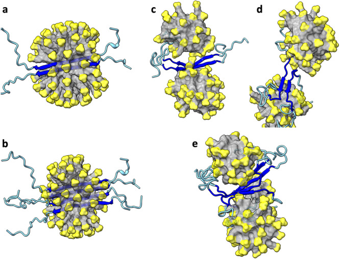
a Initial structure of dimer. b Initial structure of tetramer. c Dimer at 10 ns of a simulation. d Dimer at 601.6 ns. e Tetramer at 939.3 ns. The SDS micelles are shown in surface representation with headgroups in yellow and hydrocarbon tails in grey. Aβ42 molecules are shown in cartoon representation with residues 1-10 and 25-42 in cyan and residues 11-24 in blue.