
Fig. 5. MD simulations of the Aβ42 C-sheet dimers in SDS micelles.
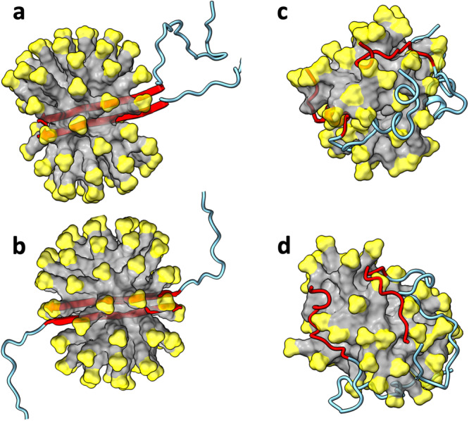
a Initial structure of parallel dimer. b Initial structure of antiparallel dimer. c Parallel dimer at 947.1 ns of a simulation. d Antiparallel dimer at 661.3 ns. The SDS micelles are shown in surface representation with headgroups in yellow and hydrocarbon tails in grey. Aβ42 molecules are shown in cartoon representation with residues 1 to 29 in cyan and residues 30-42 in red.