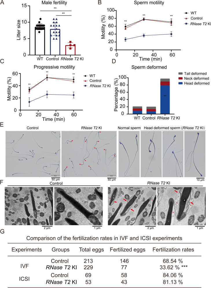
Fig. 2.
Poor male fertility and sperm quality in RNase T2 KI mice. A The litter sizes of the pregnant females showed that the RNase T2 KI males had lower fertility than WT and control males (n = 4 to 18 per group according to the number of pregnant females). B, C CASA of sperm motility showed that RNase T2 KI males display poor sperm motility (B) and progressive motility (C) in comparison to WT and control males (n = 3). D RNase T2 KI sperm exhibited significantly higher sperm head deformity (n = 16). E Wright-Giemsa staining showed head deformed sperm in RNase T2 KI mice. Scale bar, 50 μm. F Electron microscopic analysis showed intumescent cytoplasm (see the indicated asterisks) and displaced acrosome (see the indicated arrows) in sperm head of RNase T2 KI sperm. Scale bar, 2 μm and 1 μm respectively. G Comparison of the fertilization rates in IVF and ICSI experiments. Data are expressed as means ± SD; *P < 0.05, **P < 0.01 and ***P < 0.001

