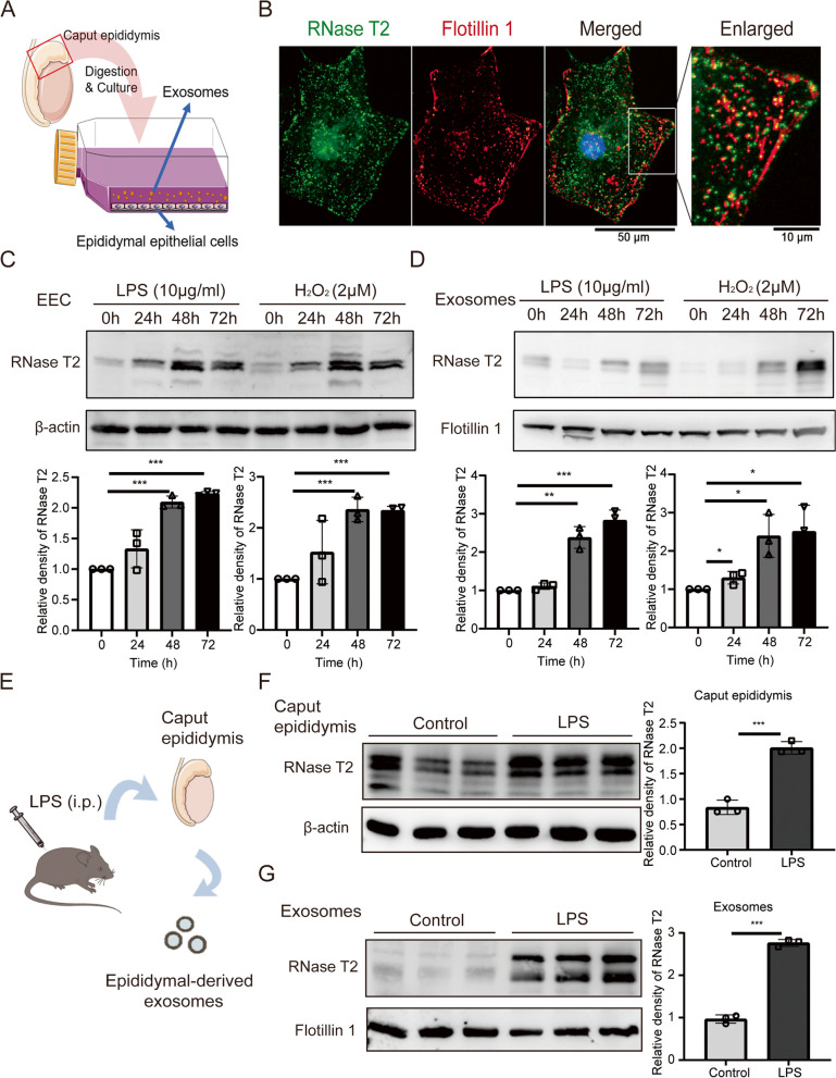
Fig. 6.
Stress conditions induce upregulation of RNase T2 in epididymal epithelial cells. A Schematic of primary culture of EEC and isolation of exosomes in the medium. B Immunofluorescent staining of RNase T2 (green fluorescence) and Flotillin 1 (red fluorescence) showed their colocalization in EEC. Scale bar, 50 and 10 μm. C, D Western blot analysis of RNase T2 in EEC (C) and isolated exosomes (D) demonstration of upregulation of RNase T2 in the presence of LPS and H2O2. Quantification of Western blots, normalized to β-actin (C, n = 3) and Flotillin 1 (D, n = 3). E Schematic representation of the isolation of epididymal-derived exosomes from inflammatory mice. F, G Western blot analysis of RNase T2 in caput epididymis and the exosomes showed the increase of RNase T2 expression in caput epididymis (F) and exosomes (G) from mice with LPS treatment. Quantification of Western blots, normalized to β-actin (F, n = 3) and Flotillin 1 (G, n = 3). Data are expressed as means ± SD; *P < 0.05, **P < 0.01 and ***P < 0.001

