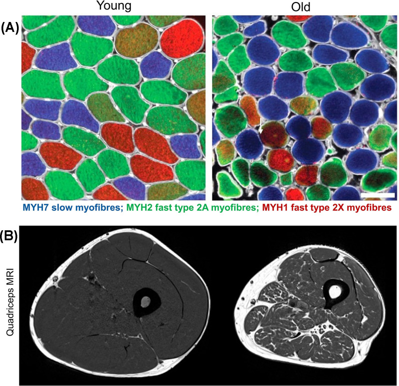
Figure 2. Comparison of skeletal muscle morphology and architecture in young and old adults.
Panel (A) depicts representative cross-sections of the vastus lateralis (VL) muscle biopsies from young (left; aged ∼24 years) and old (right; aged ∼70 years) healthy, active men, immuno-stained with antibodies specific to adult myosin heavy chain (MYH) isoforms: blue (anti-MYH7, slow type 1 myofibres), green (anti-MYH2, fast type 2A myofibres), and red (anti-MYH1, fast type 2X myofibres) (scale bar: 100 μm). Compared with the relatively uniform size of myofibres in young muscles (left; Panel A), the main features of old muscle are a wider range (variability) of size and shapes especially in type 2 myofibres (right; Panel (A), white arrow). Panel (B) shows a magnetic resonance imaging (MRI) of human thigh with area of quadriceps muscle (grey) and surrounding fatty tissue (white) in young (left) compared with loss of muscle mass in older men (right).
Panel (A) from [38] was used with the permission from Cell Press. Panel (B) from Herrmann, M., Engelke, K., Ebert, R., Müller-Deubert, S., Rudert, M., Ziouti, F. et al. (2020) Interactions between Muscle and Bone-Where Physics Meets Biology. Biomolecules10, 432, with permission from MDPI.

