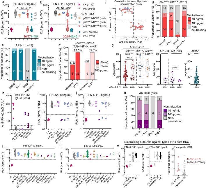
Extended Data Fig. 7. AAN-I-IFNs in patients with inborn errors of the alternative NF-κB pathway.
(a-b) Luciferase-based neutralization assay for detecting auto-Abs neutralizing 10 ng/mL IFN-α2 (a) or IFN-ω (b) in patients with the three inborn errors of NF-κB2, APS-1 and PAD patients, positive controls (C+), and healthy controls (HC). (c) Correlation between the detection of auto-Abs against IFN-α2 by Gyros (x-axis) and results for the luciferase-based neutralization assay (y-axis) after stimulation with 10 ng/mL IFN-α2. The dotted line represents the cutoffs for detection (A.U. value > 50) or neutralization (induction <5). (d-e) Proportion of patients with auto-Abs neutralizing type I IFNs at 10 ng/mL or 100 pg/mL among patients with a p52LOF/IκBδGOF variant (d), and APS-1 patients (e). (f) Proportion of patients with AAN-I-IFNs among patients carrying a missense or pLOF p52LOF/IκBδGOF variant. (g) Age distribution of patients with the three inborn errors of NF-κB2, AR RelB or NIK deficiency, or APS-1 according to the presence or absence of AAN-I-IFNs in plasma. (h) Detection of IgG auto-Abs against IFN-α2 by Gyros in patients with inborn errors of NIK, RelB, BAFF and CD40L. (i-j) Luciferase-based neutralization assay for detecting auto-Abs neutralizing 10 ng/mL IFN-α2 (i) or IFN-ω (j) in patients with inborn errors of NIK, RelB, BAFF and CD40L. (k) Proportion of patients with auto-Abs neutralizing type I IFNs at 10 ng/mL or 100 pg/mL in patients with AR RelB deficiency. (l-m) Luciferase-based neutralization assay for detecting auto-Abs neutralizing 100 pg/mL IFN-α2 (l) or IFN-ω (m) in patients with inborn errors of the canonical NF-κB pathway. DN = dominant-negative. (n) Detection of auto-Abs neutralizing 100 pg/mL IFN-α2 or IFN-ω in patients with inborn errors of the alternative NF-κB pathway post-HSCT (n = 7) versus children with inborn errors of T-cell intrinsic immunity [(SCID, n = 3, CID, n = 1), neutrophil-intrinsic immunity (chronic granulomatous disease, CGD, n = 10), cytotoxicity (familial hemophagocytic lymphohistiocytosis, HLH, n = 3), erythrocyte function (β-thalassaemia, n = 3)] who underwent HSCT (Hematop. IE, n = 20) (left panel), with the time interval between HSCT and plasma collection (right panel).

