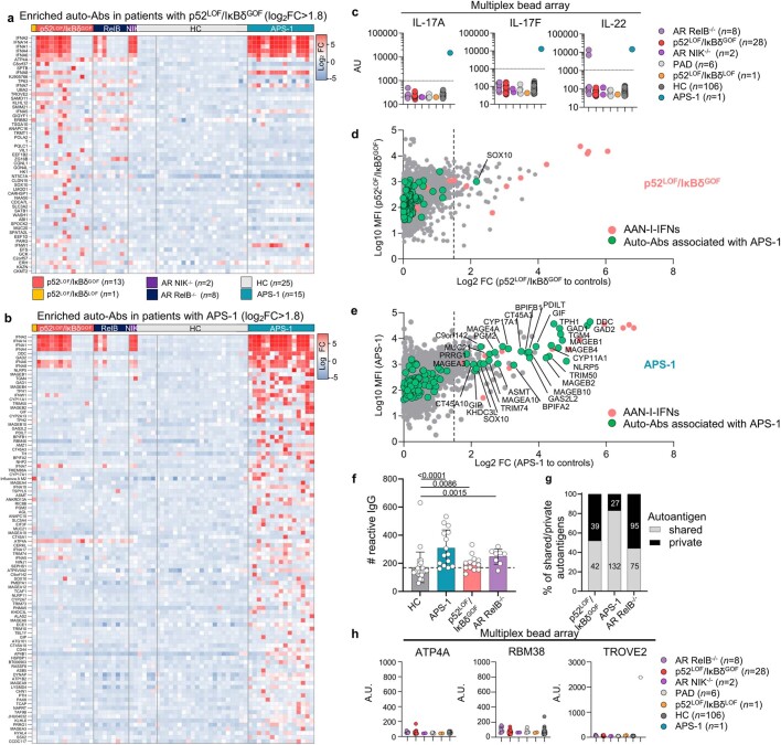
Extended Data Fig. 8. Narrow autoantibody profiles in patients with p52LOF/IκBδGOF variants.
(a-b) Heat map of the autoantigens with the highest levels of enrichment in patients with a p52LOF/IκBδGOF variant (n = 13, a) or APS-1 patients (n = 15, b), versus patients with AR RelB deficiency (n = 8), AR NIK (n = 2) deficiency, or with APS-1 (n = 15), as determined with protein microarray (HuProt). Results are shown as the mean fluorescence of two technical replicates with a log2 fold-change >1.8 in patients with p52LOF/IκBδGOF variants (a) or APS-1 (b) relative to 25 healthy controls (HC). (c) Detection of IgG auto-Abs against IL-17A, IL-17F, or IL-22 using a multiplex bead array in patients with inborn errors of the alternative NF-κB pathway. Data representative of one independent experiment are shown. (d) Protein microarray distribution of auto-Abs against IFN-α and IFN-ω (red dots) or other autoantigens frequently found targeted in patients with APS-1 (green dots), in patients with a p52LOF/IκBδGOF variant, relative to controls. (e) Protein microarray distribution of auto-Abs against IFN-α and IFN-ω (red dots), or other autoantigens associated with APS-1 (green dots) in APS-1 patients relative to controls. (f) Number of autoreactive IgG in each patient (APS-1, p52LOF/IκBδGOF, RelB−/−) or control, as determined by the sum of autoantigens with a log2 FC > 1.5 relative to the mean value for all healthy controls (HC). The error bars represent the median ± s.d. of the autoreactive IgG in each group. Comparisons done using two-tailed Mann–Whitney test. (g) Proportion of shared (by ≥ 2 patients) and private reactive autoantigens in the group of patients with a p52LOF/IκBδGOF variant, APS-1, or AR RelB deficiency. (h) Detection of auto-Abs against ATP4A, RBM38, or TROVE2 in a multiplex bead array. The white dot indicates the positive control for the detection of anti-TROVE2 auto-Abs. A.U. corresponds to arbitrary units. Data representative of one independent experiment are shown.

