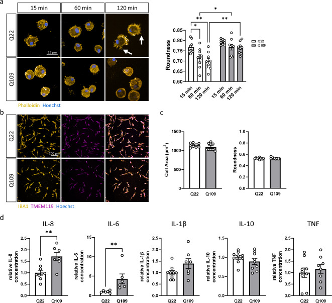
Figure 2.
Q109 iPSC differentiated to iPSC-microglia and showed no morphological differences to Q22 control iPSC-microglia but exhibit impaired attachment to extracellular matrix and increased cytokine secretion. (a) Investigation of MPC attachment to the extracellular matrix protein fibronectin 15 min, 60 min and 120 min after plating using phalloidin staining. Arrows indicate finely branched filopodia like protrusions. Roundness of Q22 MPC decreased significantly over time. This decrease was not detected in the Q109 MPC. There is no difference in roundness between Q109 and Q22 MPC 15 min after plating, but at 60 min and 120 min. Scale bar = 25 μm. (b) Confirmation of cell identity of both Q22 and Q109 iPSC-microglia using IBA1 and TMEM119. Scale bar = 100 μm. (c) Investigation of iPSC-microglia morphology showed no differences between Q22 and Q109 iPSC-microglia in their cell area and roundness. (d) Unstimulated iPSC-microglia supernatants were assessed by flow cytometric analysis for the secretion of the cytokines IL-8, IL-6, IL-1β, IL-10, TNF and IL-12p70. IL-12p70 secretion was below detection level. Unstimulated Q109 iPSC-microglia showed an increased secretion of IL-8 and IL-6. No difference was detected for the secretion of IL-1β, IL-10 and TNF. Data are expressed as mean ± SEM. n = 3 independent experimental repeats with 2–3 clones. Statistical analysis was performed using two-way ANOVA and Tukey’s honest significance test or unpaired t-test and Mann–Whitney U test (for IL-6). *p < 0.05; **p < 0.01. min = minutes.

