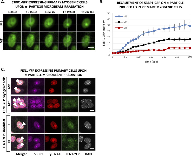
Figure S11. Decrease of 53BP1 recruitment upon α-particle microbeam irradiation in multinucleated cells is specific to myogenic cells.
(A) Sequential images of 53BP1-GFP recruitment to the locally damaged DNA (LD) by α-particle microbeam irradiation in transiently 53BP1-GFP–expressing primary myoblasts (MB, upper panel) isolated from 4–6 d old mice and subsequent differentiated myotubes (MT, lower panel). The damaged areas are underlined by a dotted cross in the nucleus. Scale bar, 10 μm. (B) Recruitment curve of 53BP1-GFP onto the LD by α-particle microbeam irradiation in transiently 53BP1-GFP transfected primary MB (blue curve), differentiating in myocytes (MC, black curve) and differentiated in MT (red curve). The irradiation was applied at t = 10 s. N ≥ 3 independent experiments with 19–26 nuclei/cell type, mean ± SEM. (C) Representative images of primary MB and MT cells (upper panel), and mononuclear (F) and fused fibroblast (FF) (lower panel), isolated from FEN1-YFP mouse model, at the indicated time post α-particle microbeam irradiation. The cells are immunolabelled with antibodies against 53BP1 (violet) and γH2AX (red). DNA was stained with DAPI (grey), and local DNA damage site is marked with an arrow head in the merged images. Scale bar, 10 μm.

