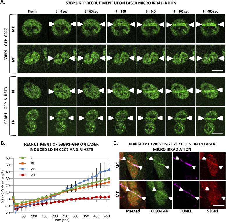
Figure S13. Almost no response of 53BP1 at local DNA damage sites induced by laser irradiation is specific to multinuclear myotubes.
(A) Sequential imaging of 53BP1-GFP recruitment to the locally damaged DNA (LD) by laser irradiation in stably 53BP1-GFP expressing C2C7 myoblasts (MB) and myotubes (MT) (upper panel) or in transiently 53BP1-GFP expressing proliferative (N) and PEG-fused (FN) NIH-3T3 cells (lower panel). The DNA damage site is indicated by two white arrow heads. Scale bar, 10 μm. (B) Recruitment curve of 53BP1-GFP to the LD by laser irradiation in stably 53BP1-GFP–expressing C2C7 MB and MT (respectively, blue and red curves) or in transiently 53BP1-GFP–expressing proliferative (N) and PEG-fused (FN) NIH-3T3 cells (respectively, green and orange curves). The irradiation was applied at t = 10 s. N ≥ 3 independent experiments with 20–34 nuclei/cell type), mean ± SEM. (C) Representative images of stably KU80-GFP expressing C2C7 myocytes (MC, upper panel) and MT (lower panel) at the indicated time post-induced local DNA damage by laser irradiation. Cells are immunolabelled with antibodies against the double-strand break markers 53BP1 (red). TUNEL labelling is used to reveal double-strand breaks (violet). Induced DNA damage site is indicated by two white arrow heads. Scale bar, 10 μm.

