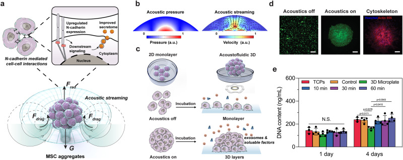
Fig. 1. Schematic of the improved stem cell secretome via acoustofluidic mechanobiological assembly.
a Schematic of the formation of MSC aggregates via acoustofluidic assembly and N-cadherin mediated cell–cell interactions to enhance the MSC secretome. b The simulation results showed that a three-dimensional circular vortex tube generated by a high-intensity focal point at the center of the substrate functioned as a virtual wall, trapping and aggregating cells at the center of the droplet. c Illustration of the comparison between 2D monolayer MSC culture and acoustofluidic 3D MSC aggregation. d Fluorescence images of MSCs before/after the acoustofluidic assembly, and the morphology of MSC aggregates after 3 days of incubation. The green color indicates alive cells. The blue color indicates cell nuclei, and the red color indicates the cytoskeleton. n = 4 tests with similar results. Scale bar: 100 μm. e CyQUANT™ Cell Proliferation Assay measuring the DNA contents of different groups. The statistical analysis was performed using one-way ANOVA with Tukey’s post-hoc test. Data are graphed as the mean ± SD (n = 4, biological repeats). Source data are provided as a Source Data file.

