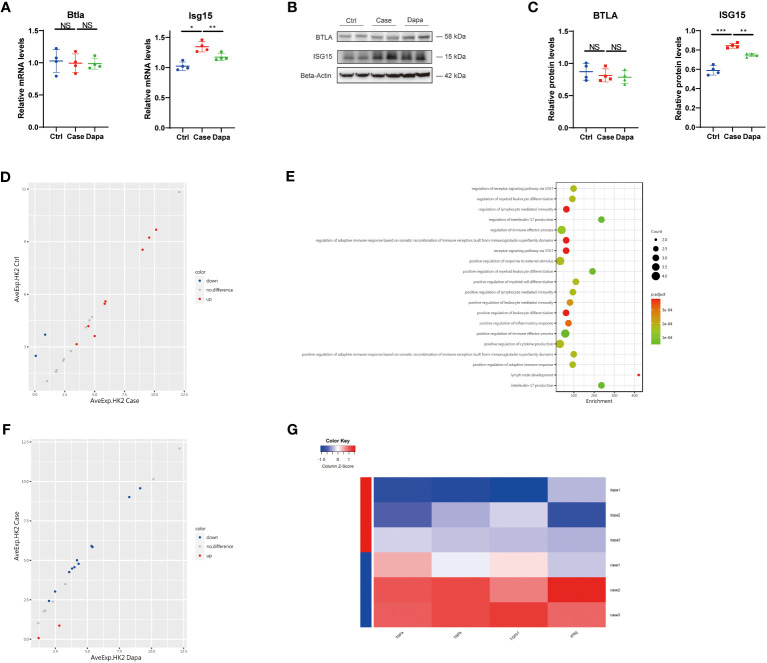
Figure 7.
Dapagliflozin presented an anti-inflammatory and anti-fibrotic effect on human renal tubular cells independent of glucose concentrations in vitro. (A) QPCR analysis showing Btla and Isg15 mRNA levels in HK-2 cells cultured in normal-glucose (Ctrl), high-glucose (Case), and high-glucose with dapagliflozin (Dapa) medium. (B) Western blot analysis showing BTLA and ISG15 protein levels in HK-2 cells of Ctrl, Case, and Dapa groups, with corresponding quantifications in (C). (D) Scatter plot indicating distinct protein expression levels of HK-2 cells in Ctrl versus Case group. (E) GO enrichment analysis suggesting STAT pathway activation, inflammatory response, and leukocyte activation pathways in HK-2 cells in the high-glucose medium. (F) Scatter plot indicating distinct protein expression levels of HK-2 cells in Case versus Dapa group. (G) Heatmap showing different protein levels of inflammatory and fibrotic cytokines in HK-2 cells of Case and Dapa group. NS, not significant. *P < 0.05, **P < 0.01, ***P < 0.001, one-way ANOVA test.

