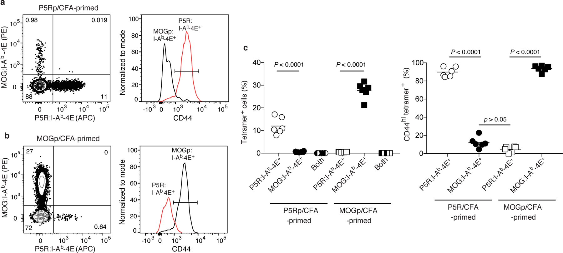
Fig. 3 |. Detection of polyclonal CD4+ effector T cells p:I-Ab-4E tetramers is peptide-specific.

a, b, Representative flow cytometry contour plots of tetramer staining (left) and histograms of CD44 expression (right) by CD4+ T cells in split samples from P5R:I-Ab-4E and MOGp:I-Ab-4E tetramer-enriched samples of spleen and lymph nodes from B6 mice immunized with P5R (a) or MOGp (b) in CFA seven days earlier. c, Scatter plots showing the percentage of CD4+ T cells (left) from individual mice (n = 6, from 2 independent experiments) detected with the indicated tetramers in tetramer-enriched samples or the percentage of CD44hi cells among the indicated tetramer-binding populations (right) with horizontal bars at the mean values. Mean values were compared with by one-way analysis of variance (ANOVA) with Tukey’s multiple comparisons test. P values are shown.
