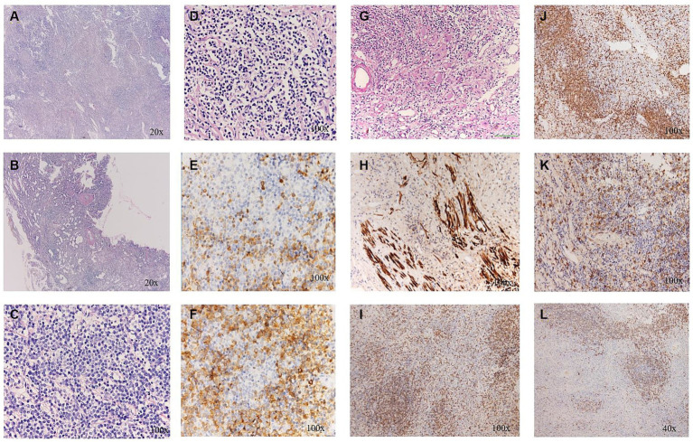
Figure 4.
(A) The distinct fibrosclerotic background; (B) Sclerosis and hyalinization of the wall of small vessels; (C) A large number of lymphocytes; (D) Patches of plasma cells; (E,F) More than 50 IgG4positive cells per high-power field. (G) Ganglion cells under H&E staining; (H) S-100 positivity; (I) CD 3(+); (J) CD 4(+); (K) CD 20(+); and (L) CD 21(+).

