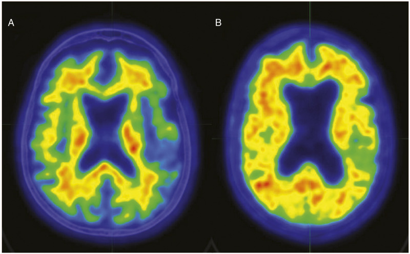
Figure 2.
Color scale (white > red > yellow > green > blue) representative 18F-Flutemetamol amyloid images for 2 participants. Participant A is an Aβ-participant (SUVR = .46); Participant B is an Aβ+ participant (SUVR = .85). Abnormal 18F-Flutemetamol uptake is indicated by loss of the normal distinct gray and white matter contrast (ie, white matter uptake greater than cortex) and areas of equal or greater uptake in the cortex in relation to white matter. Both participants were 80-year-old females with 16 years of education, and fell in the severely impaired range on memory testing (eg, first percentile for RBANS Delayed Memory Index). Participant A’s motor task performance was 62.89 seconds, while Participant B’s was 93.59 seconds. PET images follow radiological convention; both images are of CT slice 21 of 47.PET: positron emission tomography; RBANS: Repeatable Battery for the Assessment of Neuropsychological Status; SUVR: standardized uptake value ratios.

