Fig. 4.

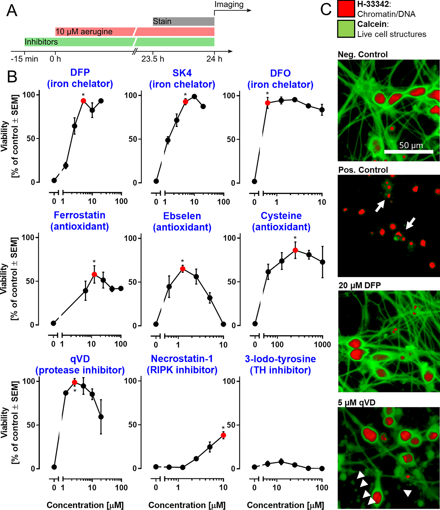
Modulation of aerugine-induced neurodegeneration by mechanistic inhibitors. LUHMES cultures (d6) were used under standard conditions to assess the cytotoxicity of aerugine (10 μM) in the presence of various agents known to interfere with cell death pathways. A Treatment scheme: the respective inhibitors were applied 15 min prior to aerugine treatment (10 μM), and the viability was determined by high-content imaging after 24 h. B Inhibitors were used at different concentrations, as indicated. The main biochemical activity of each agent is indicated in brackets. Data are means ± SEM of at least three biological replicates with 3 technical replicates each. DFP = deferiprone; DFO = deferoxamine; qVD = caspase inhibitor; RIPK = receptor-interacting serine/threonine-protein kinase. For statistical analysis, the lowest concentration of inhibitor that produced the maximal protective effect (red) was compared to the treatment with aerugine alone using a two-tailed t-test (*: p = 0.01). C Exemplary pictures of LUHMES cultures after 24 h treatment: negative control (=DMSO), positive control (=10 μM aerugine), DFP (=20 μM DFP + 10 μM aerugine) and qVD (=5 μM qVD + 10 μM aerugine). Arrows indicate cells with a broken cell membrane (calcein-negative) and condensed nuclei (small H-33342-positive area). Arrow heads indicate disintegrating neurites (chains of calcein-positive blebs). Enlarged details are given in Fig S12.
