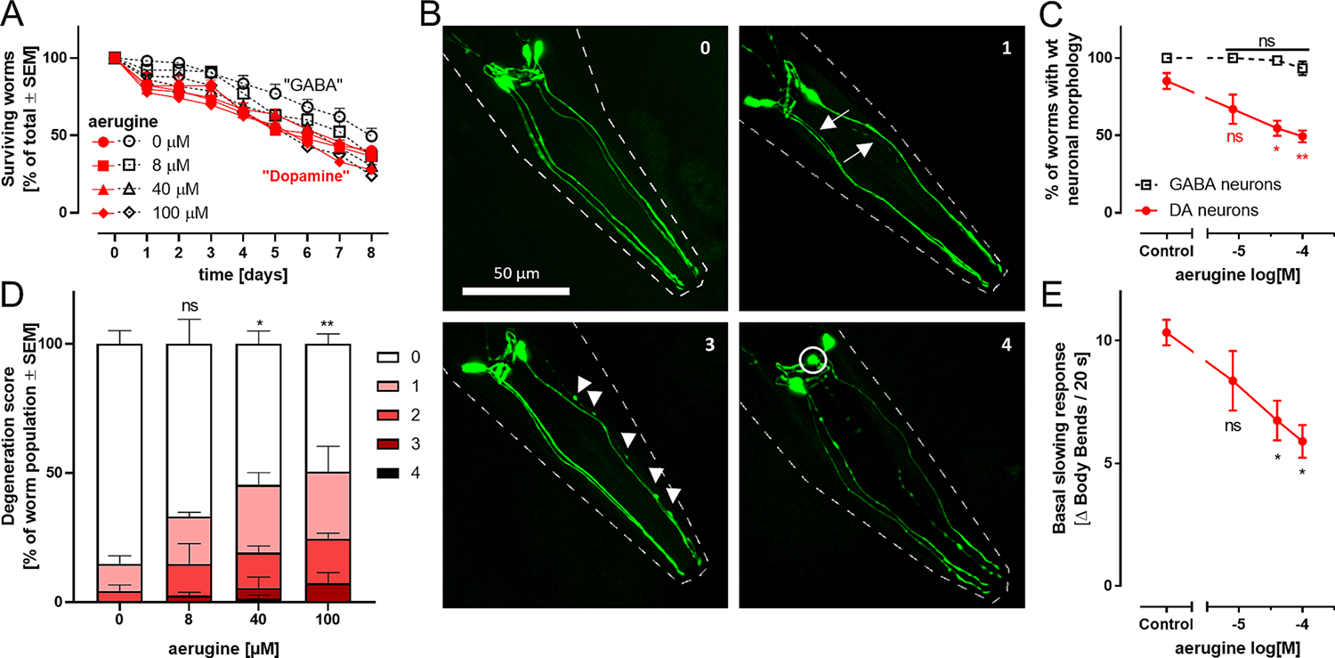
Fig. 5.

Specific dopaminergic neurodegeneration triggered by aerugine in C. elegans. A The C. elegans worm strains BY200 (containing fluorescently labelled dopamine neurons; label “Dopamine”) and Punc-25 (containing fluorescently labelled γ-aminobutyric acid neurons, label “GABA”) were used at L4 larval stage for an eight day survival test. They were exposed to different aerugine concentrations and counted every 24 h. Data are based on 40–60 worms per group. Differences on a given day were analyzed by ANOVA and found to be non-significant. Two-way ANOVA (time × treatment) indicated a significant trend of decreased survival over time (independent of treatment or genotype). B L4 larval stage worms of both strains were treated for 2 days with aerugine. The GFP-labelled neurons of 20–30 worms per condition were analyzed and scored for specific neurodegeneration. Each worm was assigned a degeneration score (0 for wild type and 1–4 for increasing severity). Representative images illustrating neurodegeneration scores (indicated by numbers in the upper right corner): The pictures show the 4 cephalic dopaminergic neurons in green. The worm outline is indicated by dashed white lines. Arrows highlight thinning dendrites. Arrow heads point out blebs (irregular protrusions of the dendrites). Degenerating cell bodies are circled. Degeneration score 2 looks similar to 3 but has a maximum of 4 blebs. These scores were assessed to quantify the dopaminergic neurodegeneration caused be aerugine in vivo. C Worms with wild type morphology (score 0) were counted. For statistical analysis, a one-way ANOVA with Dunnett’s multiple comparisons test was performed (*: p < 0.05; **: p < 0.01 for difference of aerugine groups vs untreated controls). D The degeneration scores exemplified in B were quantified. The data for DAergic neurons is shown. The data for GABAergic neurons can be found in Fig S13. E The “basal slowing response” was assessed as proxy for the functionality of the worms’ dopaminergic system. It was measured in L4 larval stage BY200 worms treated for 2 days with aerugine. As the slowing response is triggered by food, the body bends per 20 s in the absence and presence of food were counted and the difference (Δ) was calculated as experimental readout. For statistical analysis, a one-way ANOVA with Dunnett’s multiple comparisons test was performed (*: p < 0.05).
