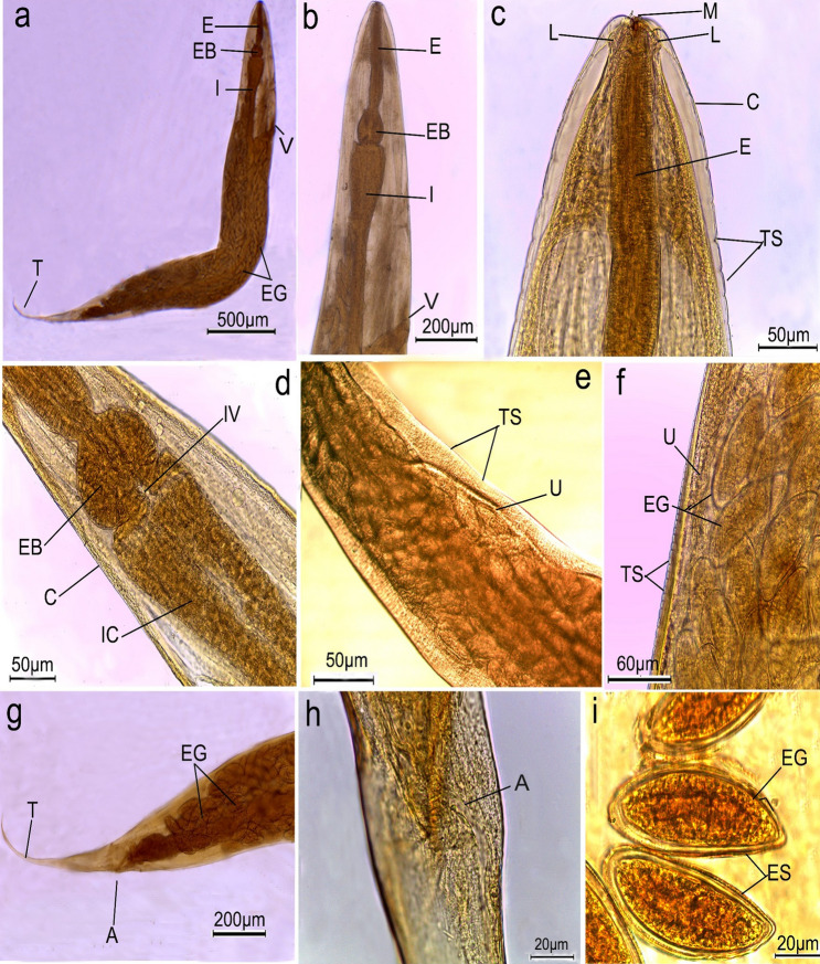
Fig. 3.
Photomicrographs of female Syphacia muris isolated from control rats cleared with lactophenol. a whole worm showing oesophagus (E), oesophageal bulb (EB), intestine (I), vulva opening (V), eggs (EG) and pointed tail (T); b, c high magnification of anterior region showing mouth (M) surrounded by three lips (L), oesophagus (E), body covered by cuticle (C) with transverse striations (TS) and vulva opening (V); d fore-body region showing oesophageal bulb (EB), intestinal valve (IV) lead to intestinal caeca (IC); e, f mid-body region showing uterus (U), folds containing eggs (EG); g, h posterior end of female worm showing anal opening (A) and terminated with tail tip (T) i high magnification of eggs (EG) surrounded by egg sheath (ES)

