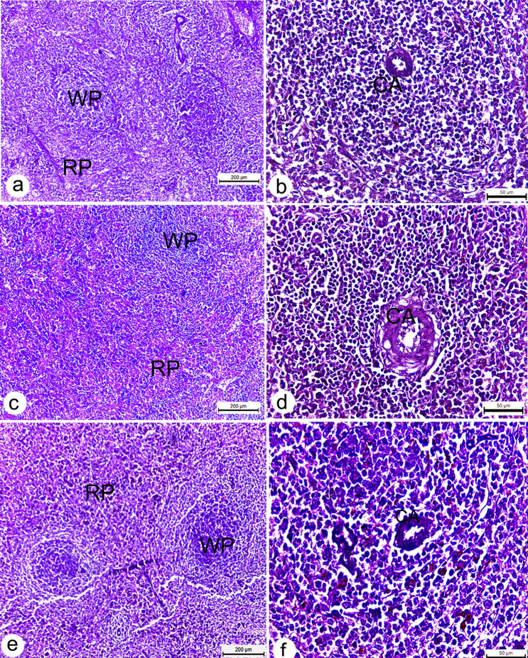
Fig. 8.
Photomicrographs of splenic sections of Wistar rats. a, b spleen section of untreated rats showing well defined splenic architecture, including healthy lymphoid cells, sinuses and central artery (CA); c–f spleen section from treated rats received 2,4 mg/kg b.w. Ag NPs showing normal histology of both red (RP) and white pulp (WP). All sections stained with H&E. scale bars a, c, e = 200 µm, b, d, f = 50 µm

