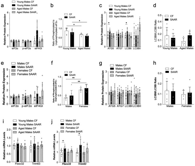
Figure 3.
The effects of SAAR on mouse muscle proteostasis is minimally affected by age of diet initiation and biological sex. Muscle tissues (quadriceps femoris) were collected at the end of diet intervention from young mice, which were fed the diet at 8 weeks old for 1 year; aged mice, which were fed the diet at 2 years old for 15 weeks; and middle-aged male and female mice, which were fed the diet at 1 year old for 52 weeks. These mice were given either a control diet (CF, 0.86% methionine w/w) or an SAAR diet (0.12% methionine w/w), as outlined in the Methods section. Proteins and RNA were extracted, and markers related to protein synthesis, autophagy, and atrophy were measured using Western blot and quantitative PCR, respectively, as outlined in the Methods section. White dots within bar graphs indicate values from each animal. Band intensities for each protein was normalized to Ponceau S stain from the same membrane from young and aged males (a and c), and middle-aged males and females (e and g). The ratios of phosphorylated to total proteins of eIF2α (b and f), and LC3BII to LC3BI (d and h) in muscles from CF and SAAR mice were quantified as described in Methods. Original membrane blots are presented in Supplementary Figure S3 and S4. Expression of atrophy related genes Fbxo32 and Trim63 from muscles of young and age (i) and middle-aged male and female mice (j) fed either a CF or SAAR diets. Comparisons between diets in the same age and sex were analyzed using Student’s t-test, as described in Methods (n = 5–8 per group, *P < 0.05, ****P < 0.0001).

