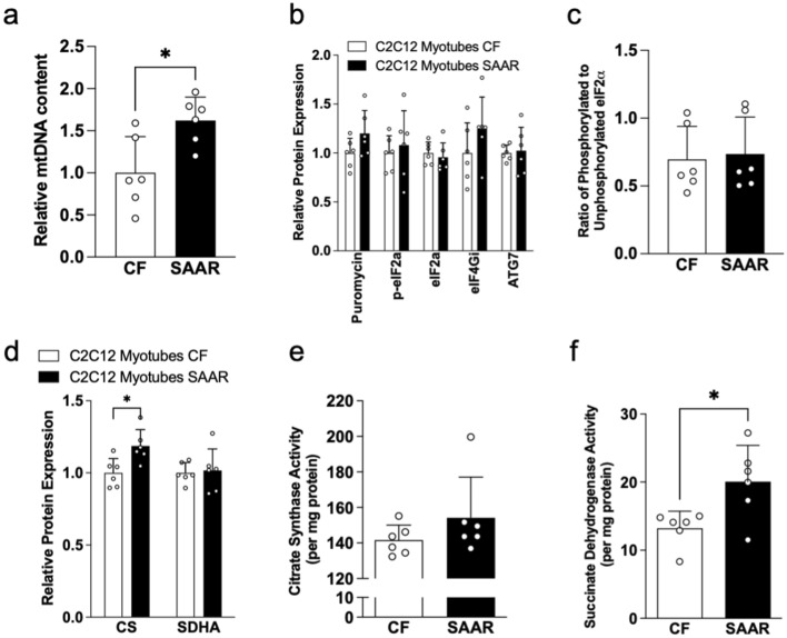
Figure 5.
SAAR enhanced mitochondrial activity in C2C12 myotubes. Relative mitochondrial DNA content in differentiated C2C12 myotubes exposed to either CF (white bars) or 80% SAAR (black bars) media for 24 h (a) as described in Methods. White dots within bar graphs indicate values from each well. Proteostasis-related protein expressions, including puromycin, phosphorylated and total eIF2a, eIF4GI, and ATG7, were assessed in C2C12 myotubes exposed to either CF or SAAR conditions as described in Methods (b). The ratio of phosphorylated to unphosphorylated eIF2a in C2C12 myotubes was also determined (c). Furthermore, protein expressions of mitochondrial activity enzymes, such as citrate synthase (CS) and succinate dehydrogenase (SDHA), were measured in C2C12 myotubes exposed to either CF or SAAR conditions as detailed in Methods (d). Whole membrane immunoblots are presented in Supplementary Figure S5. CS and SDH activities (e and f, respectively) determined by enzymatic assay described in Methods. Comparisons between treatments were analyzed using Student’s t-test, as described in Methods (n = 6 per group; *P < 0.05).

