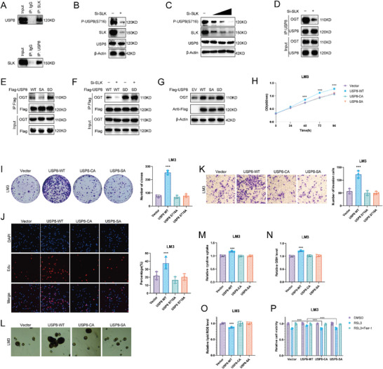
Figure 6.

SLK phosphorylates USP8. A) Co‐IP assay reveals an association between endogenous USP8 and SLK in LM3 cells. LM3 cells were harvested with RIPA lysis buffer. Co‐IP was performed using antibody as indicated. B) The expression of USP8 serine 716 phosphorylation was detected in LM3 cells. C) The expression of USP8 serine 716 phosphorylation was detected in LM3 cells transfected with increasing amounts of SLK siRNA. D) Co‐IP assay with anti‐USP8 antibody in LM3 cells treated as indicated. E) The Co‐IP analysis of OGT and Flag‐USP8 WT, S716A, or S716D in HEK293T cells. F) LM3 cells transfected with Flag‐USP8 WT or S716D mutants were treated with siControl or siSLK. G) LM3 cells were transfected with Flag‐USP8 WT, S716A, or S716D mutants, and OGT protein levels were detected. H) CCK8 assays of LM3 cells transfected with USP8 WT, S716A or C786A. I) Colony formation assays of LM3 cells transfected with USP8 WT, S716A, or C786A. J) Edu assays of LM3 cells transfected with USP8 WT, S716A, or C786A. K) Cell invasion assay of LM3 cells transfected with USP8 WT, S716A, or C786A. L) Sphere formation assay of LM3 cells transfected with USP8 WT, S716A, or C786A. M–O). Cystine, GSH, and lipid ROS levels were quantified in LM3 cells transfected with USP8 WT, S716A or C786A. P) CCK8 assay showing the response of LM3 cells to RSL3 (10 µm)±ferrostatin (1 µm). Results shown are representative of three independent experiments. Data are represented as mean ± SD of biological triplicates.*p value < 0.05; **p value < 0.01; and ***p value < 0.001.
