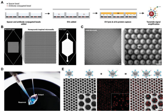
Figure 1.

Device and schematic. A) Schematic of double digital EV microwell protein detection. B) CAD files of the bottom microwell layer and top flow layer. C) PDMS microwell devices: width (W) = 42 µm and distance (D) = 20 µm. (Scale bar 50 µm) D) Loading bonded microwell device with a solution in the reservoir and washing the system with a pump via connected tubing. E) Visualization of spacer streptavidin beads and capture streptavidin beads with conjugated biotin‐NHS‐Alexa Fluor (AF) 647 linker loaded into the microwell device. (Scale bar: 50 µm)
