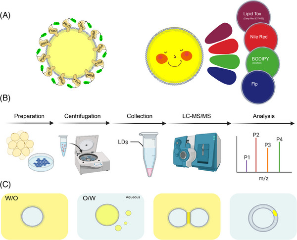
FIGURE 4.

Common methods for investigating lipid droplets (LDs). (A) Visualisation of LDs. Visualisation of LDs is generally performed by two methods. One is immunofluorescence by labelling the LD‐specific protein Plin2. Or by fluorescent dyes such as Nile Red, BODIPY and LipidTox. (B) The separation and purification of LDs requires three main steps: first, sample preparation and fragmentation; second, separation of LDs and other cellular fractions by ultracentrifugation; and finally, collection and purification followed by subsequent experiments. (C) There are two mainstream models for in vitro construction of LDs, oil‐in‐water (O/W) and water‐in‐oil (W/O). Based on these two models, there are new bilayer models for droplet interface bilayer and droplet‐embedded vesicles (DEVs).
