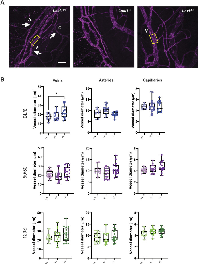
Fig. 5.
Young BL/6 Loxl1−/− mice have dilated intrascleral veins. (A) Representative images of anterior chamber flat mounts immunostained with anti-CD31 antibody in BL/6 Loxl1+/+, Loxl1+/− and Loxl1−/− mice. Yellow rectangles highlight the difference between distal vein diameter in Loxl1+/+ versus Loxl1−/− mice. A, artery; C, capillary; V, vein. Scale bar=100 µm. (B) Vessel diameter measurements for Loxl1+/+, Loxl1+/− and Loxl1−/− mice from the three different studied backgrounds. Only BL/6 Loxl1−/− mice displayed significantly dilated distal veins but not arteries or capillaries (n=4 eyes/mice, 4 quadrants/eye, *P<0.05). Boxes show the 25-75th percentiles, whiskers show the minimum and maximum, and the median is marked with a line. Ordinary one-way ANOVA followed by Dunnett's multiple comparisons test was used for statistical analysis.

