

Abstract
A pregnant woman was revealed to have fetal univentricular heart and megacystis by a routine first-trimester ultrasound. Chorionic villus sampling with the use of karyotyping and microarray found no causative etiologies. A further investigation with whole-exome sequencing (WES) demonstrated a FOXF1 variant. Autopsy confirmed the prenatal findings, and a histological study of the lungs showed the characteristic features of alveolar capillary dysplasia with misalignment of pulmonary veins (ACDMPV). This study indicates that although ultrasound itself has no ability of the identification of pulmonary histological malformations associated with ACDMPV, the early markers of univentricular heart and megacystis might alert clinicians to consider this genetic disorder which is facilitated considerably by the increasingly used WES in prenatal diagnosis.
Keywords: Alveolar capillary dysplasia with misalignment of pulmonary veins, first-trimester ultrasound screening, prenatal diagnosis
INTRODUCTION
Alveolar capillary dysplasia with misalignment of pulmonary veins (ACDMPV) is a lethal disorder, characterized histologically by failure of formation and ingrowth of alveolar capillaries.[1] Affected infants typically develop progressive, irreversible hypoxemia and respiratory failure within the first 2 days of life and die in the neonatal period. The diagnosis is generally established after lung biopsy or postmortem lung examination. Only a few cases were reported in prenatal diagnosis.[2] We report here a case of ACDMPV identified by first-trimester ultrasound, confirmed later by whole-exome sequencing (WES) and postnatal histopathology.
CASE REPORT
A 34-year-old G2P1 woman was referred to our unit at 12 weeks’ gestation because of fetal abnormality. Her husband was healthy, and they were nonconsanguineous. The family had a healthy 4-year-old daughter. The detailed first-trimester scan showed fetal megacystis with suspected hypoplastic left heart syndrome (HLHS) [Figure 1]. Chorionic villus sampling (CVS) with cell culture and microarray identified a normal female karyotype. A follow-up scan at 16 weeks found the persistence of megacystis with bilateral pyelectasis. Fetal echocardiography confirmed the diagnosis of HLHS. After genetic counseling, the stored DNA obtained from the CVS combined with parental blood samples was sent for trio-WES. This revealed a de novo heterozygous variant NM_001451(FOXF1):C. 693_694insGCGGCGCG (p.A232Rfs*150) in the fetus, confirmed by Sanger sequencing. This variant was classified as likely pathogenic according to the American College of Medical Genetics and Genomics criterion.
Figure 1.
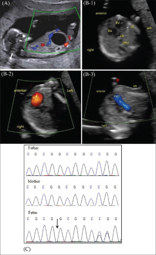
Prenatal ultrasound of the fetus. (A) Megacystis at 12 weeks; (B) Hypoplastic left heart at 12 weeks: (B-1) slit-like left ventricle and left atrium; (B-2) right atrioventricular blood flow pattern, with no flow through the left side; (B-3) only duct visualized and no V-shaped configuration); (C) Chromatograms of the FOXF1 variant by Sanger sequencing in the family members. Dao: Descending aorta; LV: Left ventricle; R: Right; RA: Right atrium; RV: Right ventricle; RVOT: Right ventricular outflow tract
Variants of FOXF1 have been associated with ACDMPV. Although the present variant is a novel one, a similar variant p.Ala231Argfs*61 was reported in an ACDMPV patient.[3] Due to severity of fetal malformations and the presumptive diagnosis of ACDMPV, the parents opted for a termination of pregnancy at 20 weeks’ gestation at another hospital. Autopsy report obtained showed the characteristic features of alveolar capillary dysplasia with misalignment of the pulmonary veins at pseudoglandular stage in the lungs.
DISCUSSION
ACDMPV is a rare, lethal developmental disorder of the lung in neonatal period, caused by variants in FOXF1 gene.[4] This gene produces FOXF1 protein that is important in the development of pulmonary mesenchyme, from which pulmonary blood vessels arise. Variants of FOXF1 have been identified in 70%–90% of potential ACDMPV cases.[3] The clinical findings of our case fit into the spectrum of malformations previously associated with FOXF1 haploinsufficiency. Most patients have concurrent congenital nonlethal malformations, including gastrointestinal, cardiac, genitourinary, right–left asymmetry, as well as congenital diaphragmatic hernia and phocomelia.[5,6] The HLHS has been reported in ACDMPV patients. Indeed, cardiac lesions have been reported in up to 25% of children with ACDMPV, with the two predominant heart conditions being left-sided obstructive lesions and atrioventricular septal defects.[7] However, the megacystis was the first description associated with this disorder. The findings of extrapulmonary anomalies and fetopathological results argue strongly for the FOXF1 variant as the causative etiology of our case.
Prenatal diagnosis of ACDMPV is challenging since the involved pulmonary histological anomalies are undetectable with routine prenatal ultrasonography. All prenatal cases reported were those in whom fetal structural abnormalities other than pulmonary anomalies were identified by ultrasound. Only a few cases were reported in the first trimester of gestation. Puisney-Dakhli et al. presented a prenatal ACDMPV case identified by ultrasound at 14 weeks of gestation.[8] The fetus had sonographic findings of increased nuchal translucency (NT) and univentricular cardiac anomaly. CVS using both microarray and fluorescence in situ hybridization detected a de novo 1.17-Mb deletion in 16q24.1 encompassing FOXF1. In the present study, we first reported the megacystis combined with cardiac anomaly as the first-trimester features which led to the prenatal diagnosis of ACDMPV. The first-trimester scan has become one of the core examinations in obstetric care, and there are studies suggesting that at least 50% of fetal major structural defects can be detected at the time of NT measurements.[9] Although these sonographic findings are nonspecific for diagnosis of a genetic syndrome, clinicians should be alert to the diagnosis of ACDMPV when extrapulmonary anomalies commonly found in ACDMPV are detected prenatally. In this case, an abnormal result of FOXF1 detected with rapid WES following a negative array is important in guiding clinical management, permitting early termination, or avoiding unnecessary deep interventions including the use of extracorporeal membrane oxygenation support.
In conclusion, we report here a prenatal case of ACDMPV diagnosed in the first trimester by ultrasound examination with its associated findings of congenital univentricular heart and megacystis. Although ultrasound itself has no ability of the identification of pulmonary histological malformations associated with ACDMPV, the early markers of univentricular heart and megacystis might alert clinicians to consider this genetic disorder. Indeed, WES should be an option for parents with fetal structural anomalies and a normal array result, which will facilitate the prenatal diagnosis, considering the growing utility of WES in the prenatal setting.
Declaration of patient consent
The authors certify that they have obtained appropriate patient’s consent form. In the form, the patient has given the consent for the prenatal images and other clinical information to be reported in the journal. The patient understands that the name and initial will not be published and due efforts will be made to conceal the identity, but anonymity cannot be guaranteed.
Financial support and sponsorship
Nil.
Conflicts of interest
There are no conflicts of interest.
REFERENCES
- 1.Vincent M, Karolak JA, Deutsch G, Gambin T, Popek E, Isidor B, et al. Clinical, histopathological, and molecular diagnostics in lethal lung developmental disorders. Am J Respir Crit Care Med. 2019;200:1093–101. doi: 10.1164/rccm.201903-0495TR. [DOI] [PMC free article] [PubMed] [Google Scholar]
- 2.Prothro SL, Plosa E, Markham M, Szafranski P, Stankiewicz P, Killen SA. Prenatal diagnosis of alveolar capillary dysplasia with misalignment of pulmonary veins. J Pediatr. 2016;170:317–8. doi: 10.1016/j.jpeds.2015.11.041. [DOI] [PMC free article] [PubMed] [Google Scholar]
- 3.Sen P, Yang Y, Navarro C, Silva I, Szafranski P, Kolodziejska KE, et al. Novel FOXF1 mutations in sporadic and familial cases of alveolar capillary dysplasia with misaligned pulmonary veins imply a role for its DNA binding domain. Hum Mutat. 2013;34:801–11. doi: 10.1002/humu.22313. [DOI] [PMC free article] [PubMed] [Google Scholar]
- 4.Slot E, von der Thüsen JH, van Heijst A, van Marion R, Magielsen F, Dubbink HJ, et al. Fast detection of FOXF1 variants in patients with alveolar capillary dysplasia with misalignment of pulmonary veins using targeted sequencing. Pediatr Res. 2021;89:518–25. doi: 10.1038/s41390-020-0931-5. [DOI] [PubMed] [Google Scholar]
- 5.Arreo Del Val V, Avila-Alvarez A, Schteffer LR, Santos F, Deiros L, Del Cerro MJ. Alveolar capillary dysplasia with misalignment of the pulmonary veins associated with aortic coarctation and intestinal malrotation. J Perinatol. 2014;34:795–7. doi: 10.1038/jp.2014.94. [DOI] [PubMed] [Google Scholar]
- 6.Abu-El-Haija A, Fineman J, Connolly AJ, Murali P, Judge LM, Slavotinek AM. Two patients with FOXF1 mutations with alveolar capillary dysplasia with misalignment of pulmonary veins and other malformations:Two different presentations and outcomes. Am J Med Genet A. 2018;176:2877–81. doi: 10.1002/ajmg.a.40641. [DOI] [PubMed] [Google Scholar]
- 7.Bishop NB, Stankiewicz P, Steinhorn RH. Alveolar capillary dysplasia. Am J Respir Crit Care Med. 2011;184:172–9. doi: 10.1164/rccm.201010-1697CI. [DOI] [PMC free article] [PubMed] [Google Scholar]
- 8.Puisney-Dakhli C, Gubana F, Petit F, Bouchghoul H, Gautier V, Martinovic J, et al. Early prenatal diagnosis of alveolar capillary dysplasia with misalignment of pulmonary veins due to a 16q24.1 deletion. Am J Med Genet A. 2021;185:1494–7. doi: 10.1002/ajmg.a.62105. [DOI] [PubMed] [Google Scholar]
- 9.Karim JN, Roberts NW, Salomon LJ, Papageorghiou AT. Systematic review of first-trimester ultrasound screening for detection of fetal structural anomalies and factors that affect screening performance. Ultrasound Obstet Gynecol. 2017;50:429–41. doi: 10.1002/uog.17246. [DOI] [PubMed] [Google Scholar]