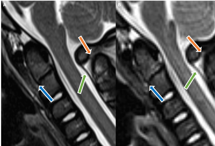
Figure 2. Sagittal T2-weighted (A) and STIR (B) MR images show abnormal hyperintensity with associated anterior tilting of the dens (blue arrows) consistent with odontocentral synchondrosal injury. Associated craniocervical junction posterior element injury involving the posterior atlantoaxial ligament (green arrows) and C1-C2 interspinous ligament (orange arrows) hyperintensity.
STIR - short tau inversion recovery, MR - magnetic resonance

