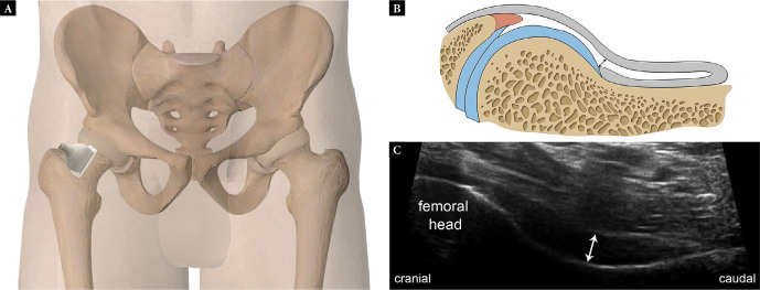
Fig. 1.
Normal anterior joint recess. Graphic illustrations (A, B) showing the transducer position (A) and equivalent anatomy (B) for long-axis scanning of the anterior hip joint recess. Long-axis US image (C) is depicted. Double-headed arrows in B and C demonstrate measurement of the recess (US image shows a recess measuring 5 mm)

