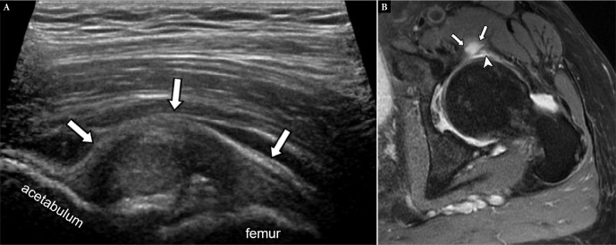
Fig. 8.
Anterosuperior paralabral cyst in a 70-year-old female with left hip pain of 4 months’ duration. Long-axis US image at the level of the hip (A) shows a complex lesion (arrows) arising from the joint space. This was noncompressible on transducer pressure, raising the possibility of a paralabral cyst. Axial PD-weighted fat-suppressed MR image (B) confirms a paralabral cyst (arrows) associated with an underlying labral tear (arrowhead)

