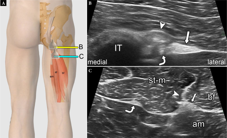
Fig. 20.
Normal proximal hamstring tendons on US. Graphic illustration (A) showing normal transducer positions and equivalent short-axis US images at the level of the ischial tuberosity (IT) (B) and proximal thigh (C) (arrowheads – conjoint tendon; arrows – sciatic nerve; curved arrows – semimembranosus tendon; st-m – semitendinosus muscle; am – adductor magnus muscle; bf – biceps femoris tendon). Fig. 20 A is modified with permission from Flores et al.(3)

