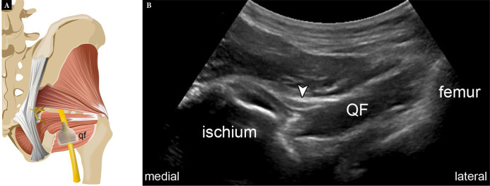
Fig. 21.
Normal ischiofemoral space. Graphic illustration (A) demonstrates normal transducer position for assessing the ischiofemoral space. Short-axis US image (B) shows the normal sonographic appearance of the space (qf – quadratus femoris; arrowhead – sciatic nerve) Fig. 21 A is modified with permission from Flores et al.(3)

