Fig. 5.
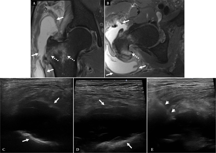
Cellulitis, bursitis, and diagnostic aspiration. 56-year-old male with severe hip pain and fever. A. Coronal and B. axial T2-weighted fat-saturated magnetic resonance (MR) images of the hip show a large, complex fluid collection (arrows) in the trochanteric bursa. Bone marrow edema is present in the adjacent femur (dashed arrows). C. Transverse and D. long-axis grayscale US images show a cobblestoned, echogenic appearance of the subcutaneous fat, consistent with edema, suggesting cellulitis. Fluid collection (arrows) is seen adjacent to the greater trochanter femoral cortex. E. US-guided aspiration was performed to confirm septic bursitis. Note needle (short arrows)

