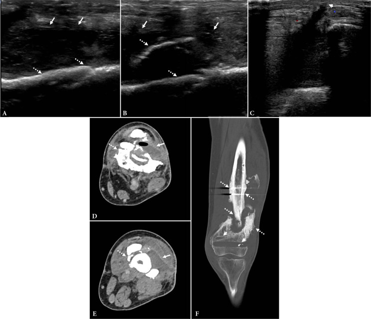
Fig. 12.
Chronic osteomyelitis. 30-year-old male previously treated for osteomyelitis involving ununited left femur fracture with multiple prior debridements and hardware removal. The patient did not complete full course of antibiotic therapy. He presented with new pain following trauma and was found to have acute on chronic osteomyelitis. A. and B. Long-axis grayscale US images show a heterogeneous hypoechoic fluid collection (arrows) adjacent to the irregular cortex (dashed arrows) from the ununited fracture. C. Transverse grayscale US image shows communication of the fluid collection to the skin surface (short arrow). D. and E. Axial contrast-enhanced CT images in soft tissue windows in the same region show chronic ununited fracture fragments (dashed arrows) as well as the peripherally enhancing fluid collection (arrows), extending from the skin surface into the fracture cavity. Note the gas in the abscess. F. Coronal reformatted CT image with bone algorithm shows sclerotic bone (dashed arrows) in the region of the fracture, consistent with chronic osteomyelitis. Note the tracts from prior hardware (short arrows)

