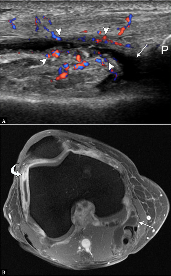
Fig. 2.

57-year-old female with osteoarthritis and synovitis. A. Longitudinal color Doppler US image superior to the patella (P) demonstrates small suprapatellar joint effusion (arrow) with hyperemic, thickened peripheral synovium (arrowheads). B. Post-contrast axial spoiled gradient echo MR image of the knee shows thickened and enhancing synovium, most prominent in the lateral gutter (curved arrow), compared to normal synovial enhancement of a Baker’s cyst (arrow). P – patella
