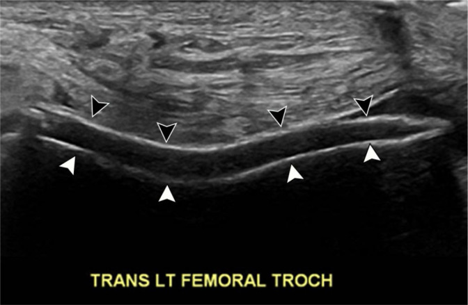
Fig. 3.

76-year-old male with acute-onset left knee pain and history of gout. Transverse grayscale US image at the level of the femoral trochlea demonstrates smooth hyperechoic layer (black arrowheads) overlying the hypoechoic hyaline articular cartilage and paralleling the hyperechoic subchondral bone (white arrowheads), consistent with layering uric acid crystals
