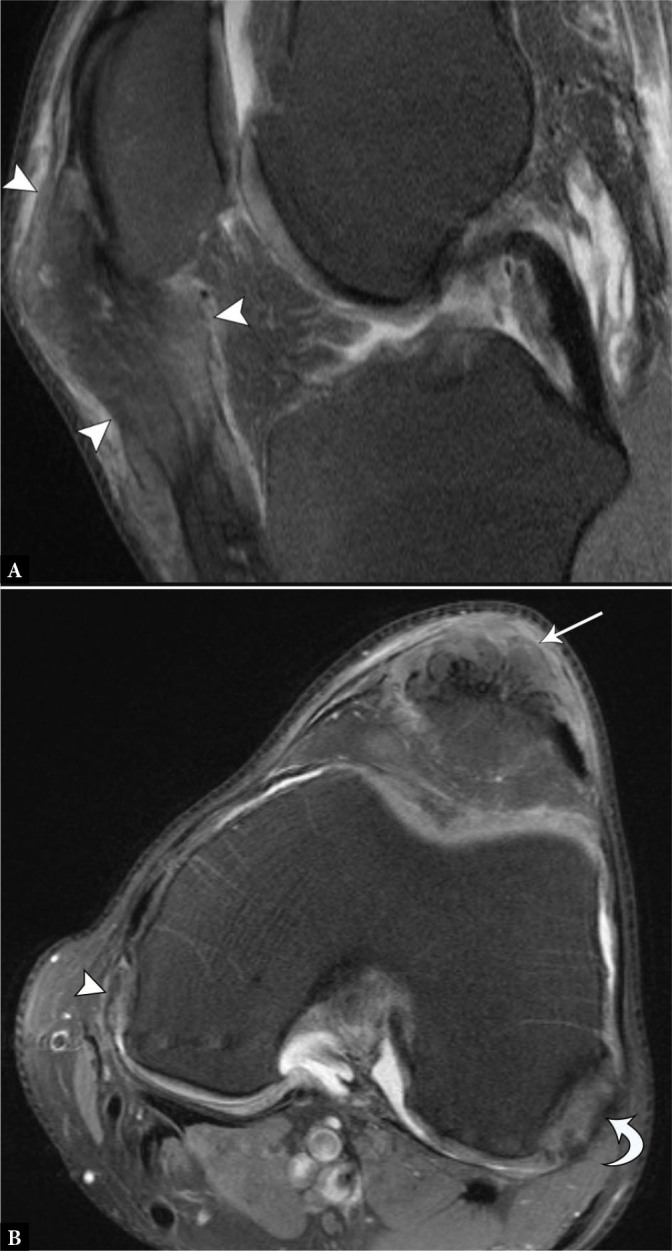
Fig. 4.

39-year-old male with tophaceous gout presenting as a palpable peri-patellar mass. A. Sagittal proton-density-weighted fat-suppressed MR image shows expansile intermediate-signal soft tissue (arrowheads) within the patellar tendon, extending superficial to the anterior patella. B. Axial proton-density-weighted fat-suppressed image shows the mass in the patellar tendon (arrow), as well as additional heterogeneously intermediate-signal nodules within the popliteus tendon (curved arrow) and along the medial femoral condyle (arrowhead), in a distribution characteristic of tophaceous gout
