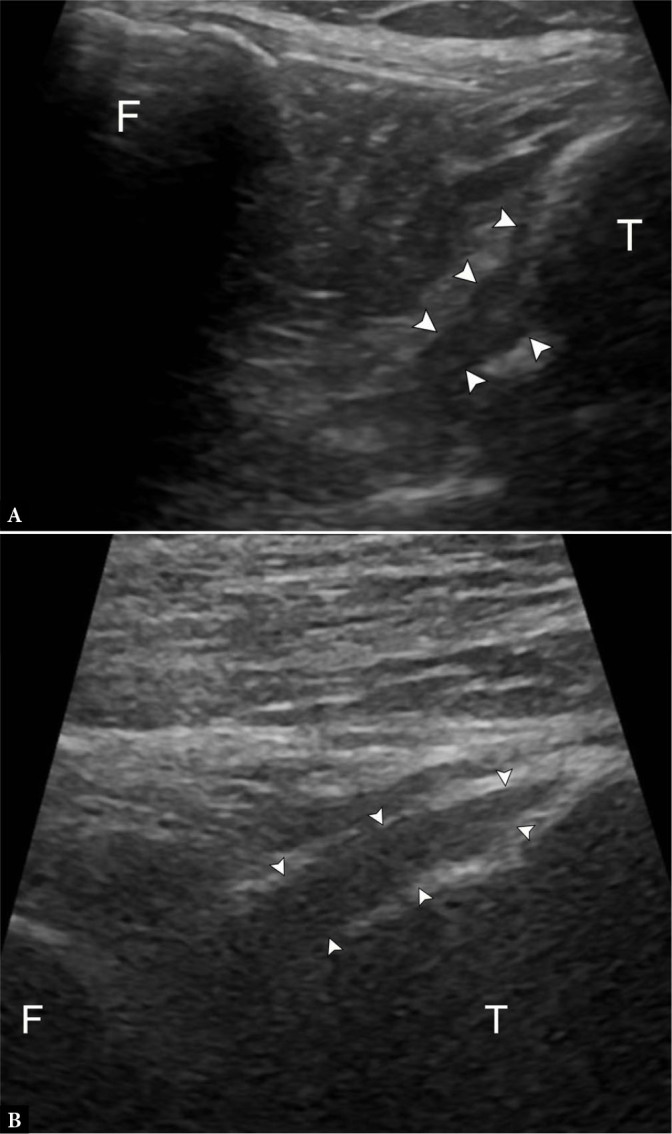
Fig. 5.

53-year-old female with knee pain, unable to undergo MRI. A. Longitudinal grayscale US image slightly oblique to the patellar tendon with knee flexion demonstrates no tear at the tibial insertion of the anterior cruciate ligament (ACL) (arrowheads). T – tibia, F – femur. B. Longitudinal grayscale US image at the central posterior tibial plateau with knee flexion demonstrates no tear at the tibial insertion of the posterior cruciate ligament (PCL) (arrowheads). The femoral origin of either ligament is not visible
