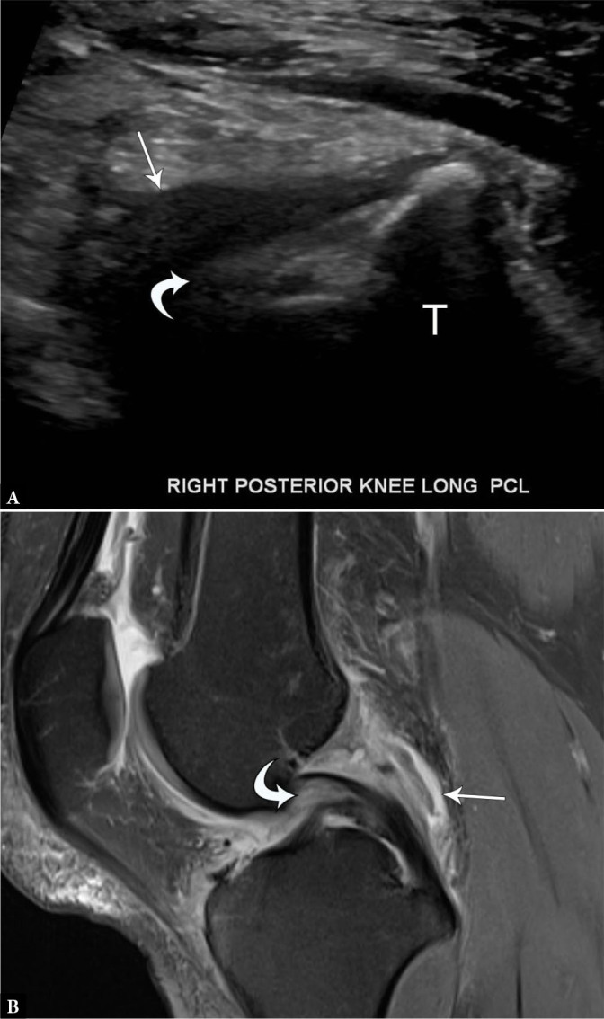
Fig. 6.

33-year-old male with partial posterior cruciate ligament tear. A. Longitudinal grayscale US image of the PCL demonstrates normal tibial attachment but indistinct, hypoechoic proximal fibers (curved arrow) with surrounding hypoechoic material (arrow). B. Sagittal proton-density-weighted fat-suppressed MR image shows edema and thickening of the proximal PCL (curved arrow) with surrounding edema and fluid (arrow)
