Fig. 8.

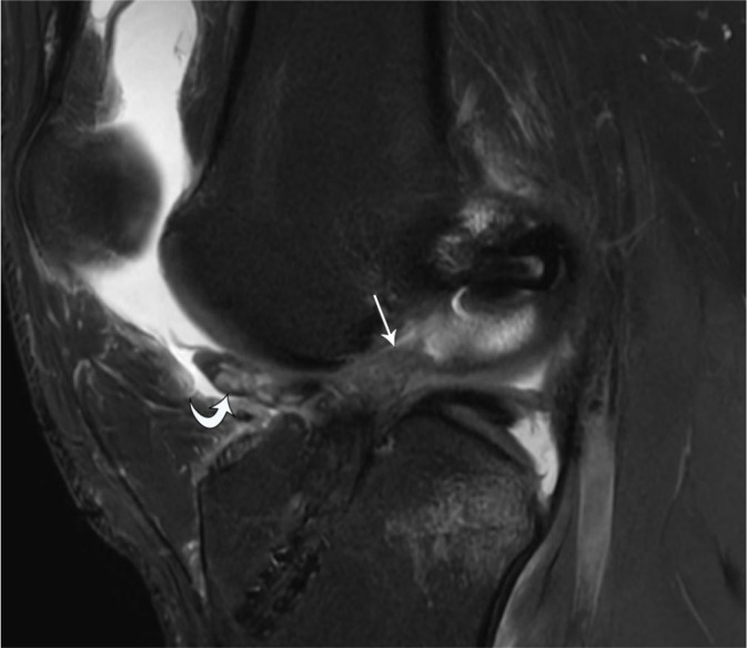
20-year-old male with complete ACL graft rupture. Sagittal protondensity-weighted fat-suppressed MR image shows complete disruption of the ACL graft (arrow) with anteriorly displaced torn graft fibers (curved arrow)

20-year-old male with complete ACL graft rupture. Sagittal protondensity-weighted fat-suppressed MR image shows complete disruption of the ACL graft (arrow) with anteriorly displaced torn graft fibers (curved arrow)