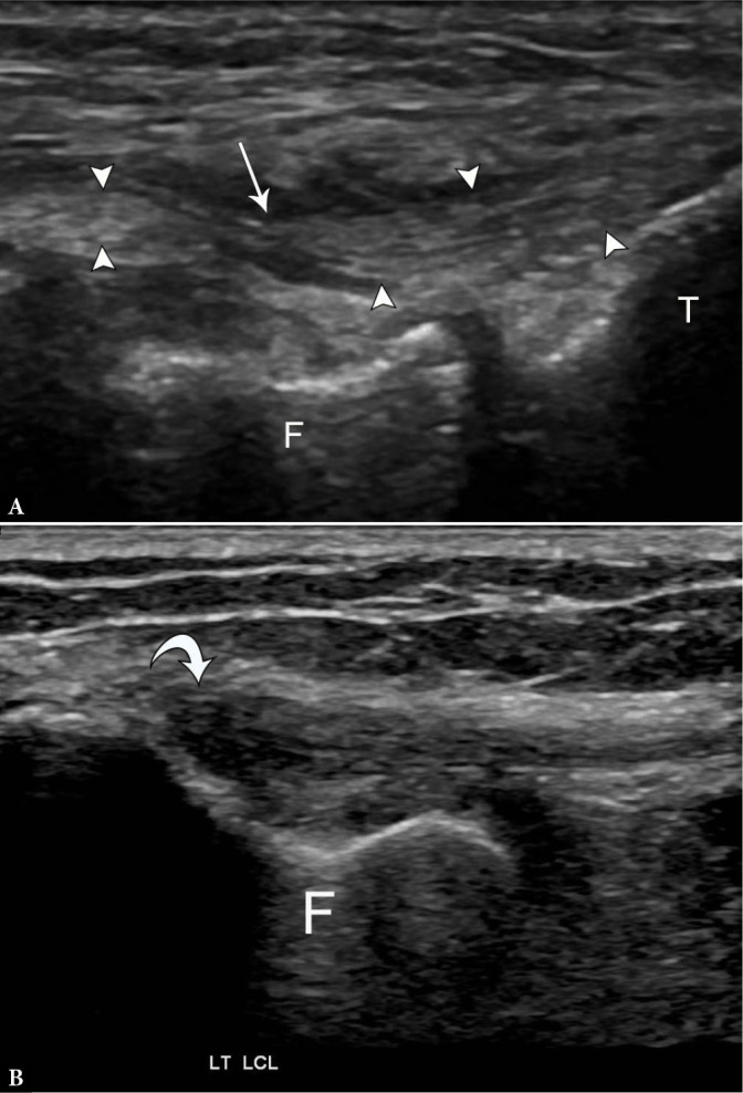
Fig. 10.

75-year-old female with high-grade partial fibular collateral ligament (FCL) injury. A. Longitudinal grayscale US image along the course of the FCL (arrowheads) next to the femoral popliteal groove (F) and tibial plateau (T) demonstrates attenuated remnant fibers with surrounding fluid (arrow). Compare to the normal appearance of the FCL on longitudinal grayscale US image B. with anisotropy (curved arrow) at the proximal portion
