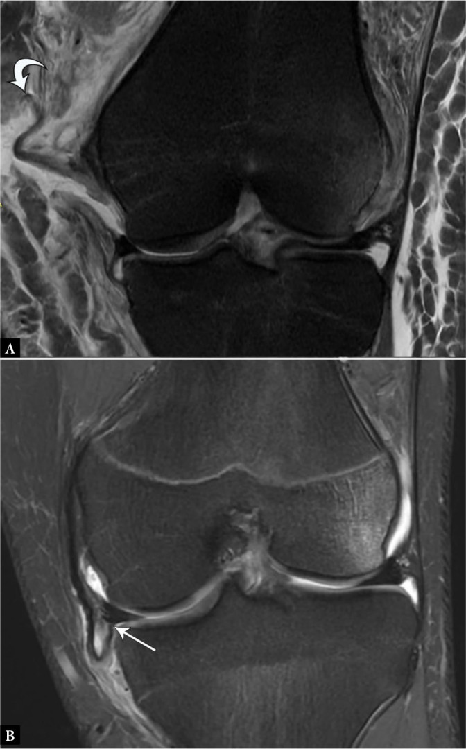
Fig. 11.

A. 19-year-old male with displaced MCL tear. Coronal proton-density-weighted fat-suppressed MR image shows completely torn and proximally displaced MCL stump (curved arrow) superficial to the pes anserinus, consistent with a Stener-like lesion. B. 16-year-old male with flipped and entrapped MCL tear. Coronal proton-density-weighted fat-suppressed MR image shows completely torn and proximally displaced MCL stump entrapped at the medial joint line inferior to the meniscus (arrow), which also requires surgical repair despite lying deep to the pes anserinus.
