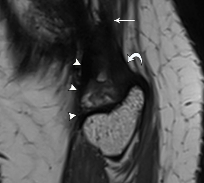
Fig. 13.

33-year-old female with snapping sensation at the lateral knee. Dynamic US imaging (see Video 1) shows snapping of the tibial arm of the biceps femoris tendon. Note initial orientation of the probe in the longitudinal plane before turning to short axis before imaging about the fibular head. Sagittal PD SPACE MR image shows thickened tibial arm (arrowheads) bifurcating from the biceps femoris tendon (arrow) and coursing anterior to the fibular arm (curved arrow)
